Laser Kidney Stone Specialist in Nashik - Dr. Pratikshit Mahajan
Dr. Pratikshit Mahajan Provide Kidney Stones Treatment In Nashik He is well known Urosurgeon In Nashik, he has lots of experience in this filed
What is Endourology?
Endourology is a minimally invasive technique available to treat kidney stones. Stones may be extracted or fragmented using tiny instruments through natural body channels such as the urethra, bladder, and ureter. In addition to treatment, our doctors help determine the cause of kidney stone development and help identify methods to prevent further stone formation. Thin, flexible instruments including lasers, graspers, miniature stone retrieval baskets, special scalpels, and cautery, can be advanced through working channels in the scopes in order to perform surgery without creating any incisions at all. The majority of endoscopic procedures can be done on an outpatient basis.
Endourological procedures include:
1. Urethroscopy: used to treat strictures or blockages in the urethra.
2. Cystoscopy: used to treat bladder stones and tumors. Obstructing prostate tissue can be removed with this approach as well (a procedure called “TURP”). Flexible plastic tubes called stents can be passed up the ureter using cystoscopy and x-rays to relieve blockage of the ureter.
3. Ureteroscopy: used to treat stones and tumors of the ureter.
4. Nephroscopy: used to treat stones and tumors of the kidney lining.
Endoscopic Procedures
Extracorporeal Shock Wave Lithotripsy
This minimally invasive technique for the disintegration of stones involves the administration of shock waves that are generated by a machine called a lithotriptor. After the machine is calibrated, and the stone has been targeted, shock waves are focused and passed through the body in such a manner that their maximum energy is dispersed at the locale of the stone, with the intent of stone disintegration. The pulverized fragments then pass in the patient’s urine. The procedure works best for smaller stones. Other determinants for success with this treatment technique include stone composition and the specific anatomic location of the stone within the urinary tract.
The University of Miami/Jackson Memorial Hospital has acquired one of the latest versions of these machines, the Lithotron Ultra. This “spark-gap” machine includes stone imaging by both x-ray and ultrasound. Experience to date has shown promising treatment results.
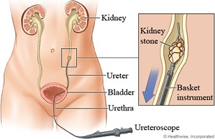
Ureteroscopy
The ureters are narrow conduits that carry urine from the kidney down to the bladder. They are normally quite small in caliber but may become dilated when they are obstructed. Ureteroscopes are precision instruments used for surgical procedures within these structures. At times a ureteroscope may also be used to traverse the length of the ureter in order to perform a procedure in the kidney.
Ureteroscopic treatment of stones
In certain cases, ureteroscopy is the most effective treatment for urinary stones. The following are specific situations where ureteroscopy is the treatment of choice:
• Where stones have not been completely broken up and cannot pass through the urine after treatment with extracorporeal shock wave lithotripsy;
• When stones are lodged in the portion of the ureter near the bladder, a region where a shock wave lithotriptor may have difficulty focusing shock waves for breakage of the stone;
• When there are stones in particular parts of the kidney (the lower portion) that even if broken up by extracorporeal shock wave lithotripsy, cannot, due to contour and angulation, pass out of the kidney; Where stones are associated with other unusual ureteral/kidney anatomy.
Various devices can be placed through the ureteroscope to facilitate stone breakage and removal. These include lasers, miniature jackhammer like stone impactors, and other similar tools that cause stones to fragment when these devices are activated. Miniature baskets can also be placed for removal of stone fragments.
The narrow ureter oftentimes swells in reaction to stone treatment, and as such, a stent, a tube resembling a thin drinking straw with curls on each end, is placed in the ureter for a few days until this swelling subsides. This stent is easily removed in the doctor’s office shortly after the procedure.
Ureteroscopic treatment of ureteral/renal strictures and other renal disorders
Due to a variety of causes, including stones that remained impacted in the ureter for a long time, the ureter may stricture; that is to say, scar and narrow to a point where urine cannot readily pass. In these situations, the obstruction of urinary flow often results in pain. However; if the obstruction is slow in onset, the patient may not notice pain specifically related to the kidney. The obstruction of urinary flow, if not relieved, will eventually result in kidney damage. These obstructions must be treated. Strictures of short length and duration can be cut open with a laser or other device placed through a ureteroscope. A stent is then placed while the ureter heals. Strictures that are longstanding and of longer length and complexity may require repeat procedures, or a more complex procedure, such as laparoscopy or open surgery, for correction.

Percutaneous Renal Surgery
Percutaneous renal surgery involves the placement of catheters through the skin in the patient’s back into the drainage system of the kidney. Subsequently, this passageway can be dilated to facilitate the placement of working tubes and instruments to break up/remove stones and to perform other necessary procedures (including relief of kidney obstruction). Though more invasive than extracorporeal shockwave lithotripsy and ureteroscopy, this procedure offers substantial benefits in terms of patient recovery when compared to open surgical procedures that, in the past, often had to be performed to treat large kidney stones or other significant kidney diseases.
Percutaneous Removal of Stones
Large kidney stones often cannot be effectively treated by extracorporeal shock wave lithotripsy or ureteroscopy. Though these procedures can be attempted, their limitations may become evident. Extracorporeal shock wave lithotripsy can be performed, but it can be difficult to break a large stone in its entirety by this means. Furthermore, the fragments that are broken must all pass, which may cause significant discomfort for the patient for a prolonged period of time. It then becomes necessary for more procedures, which require anesthesia, to be performed to remove the residual stone. Ureteroscopy can also be performed, but because the ureteroscope and the ureter are of small caliber, it may be difficult to break a large stone by this means, and then remove all of the fragments. Once again, a greater number of procedures, which require anesthesia, may need to be performed.
